High content analysis is a term often applied to image cytometry in the context of high throughput screening while tissue cytometry refers to the application of these techniques to cells in situ. This process is performed at rates of thousands of cells per second.
Analyzing Single Cells With Flow Cytometry
Filby explains how the imaging flow cytometer.

. Typical samples include everything from a cell line expressing Green Fluorescent Protein to. How a Flow Cytometer Works. Image cytometry however carries the added ability of three-dimensional imaging.
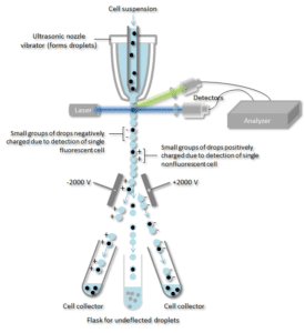
This information can be used to individually sort or separate subpopulations of cells. With chapters on instrumentation useful reagents controls experimental set up and much more this guide enables best practice to be followed and gives practical advice on building multicolor panels with example protocols. That is one way to think about it The Amnis system is an imaging flow cytometer that comes.
FCS Express Image Cytometry brings everything you would expect in a dedicated flow analysis software directly to your imaging analysis pipeline. Goda K123 Filby A4 Nitta N12. Image cytometry is generally used for the multiparameter analysis of individual adherent cells and is used to measure many of the same parameters as flow cytometry.
This technique allows researchers to get highly specific information about individual cells. Image analysis while allowing the same multiparametric capabilities as flow cytometry also permits visualization of the measured events for better quantification of less cellular specimens such as fine needle samplesIntroduction. In this talk Dr.
History Flow cytometry developed from microscopy. Cells are then loaded onto a slide and imaged using an imaging cytometer which analyzes fluorescent intensities as well as identifies individual cells. In some ways Flow cytometry IS image cytometry its just that the image resolution is only 1 pixel.
Flow cytometry has emerged as a critical component in the evaluation of primary immune deficiency disorders. In this manner cell populations can often be. IFC provides quantitative spatially registered image data for every event analyzed which allows the morphometric.
In Flow Cytometry Image Is Everything Keisuke Goda123 Andrew Filby4 Nao Nitta12 Key terms imaging flow cytometry. Further flow cytometry sacrifices imaging entirely in favor of high acquisition rates and fluorescence sensitivity. Epub 2019 May 3.
Flow cytometry is a very powerful tool for the complex characterization of cells and cell populations but flow cytometers can also be thought of as microscopes with very poor spatial resolution. As such a number of imaged-based cytometric approaches exist that can be grouped into a relatively broad field known as Imaging Cytometry. One of the fundamentals of flow cytometry is the ability to measure the properties of individual particles.
FS correlates with cell size and SS is proportional to the granularity of the cells. Andrew Filby provides an overview of imaging flow cytometry a powerful technique used to measure the phenotype of cells using image-based metrics. From plots to gates to regression analysis and high content screening see what FCS Express Image can do for you.
Flow cytometry studies are used to identify and quantify the cells of the immune system and to characterize hematological malignancies. This technique allows researchers to get highly specific information about individual cells. IFC provides quantitative spatially registered image data for every event analyzed which allows the morphometric.
Increase the resolution and you have a machine that can provide comprehensive analysis and in-depth imagery of every individual cell. With the combined advantages of optical microscopy and flow cytometry imaging flow cytometry IFC has quickly become an established tool for performing cytometric analysis in diverse areas of biology including microbiology immunology and stem cell biology 1-14. 2Japan Science and Technology Agency Kawaguchi Japan.
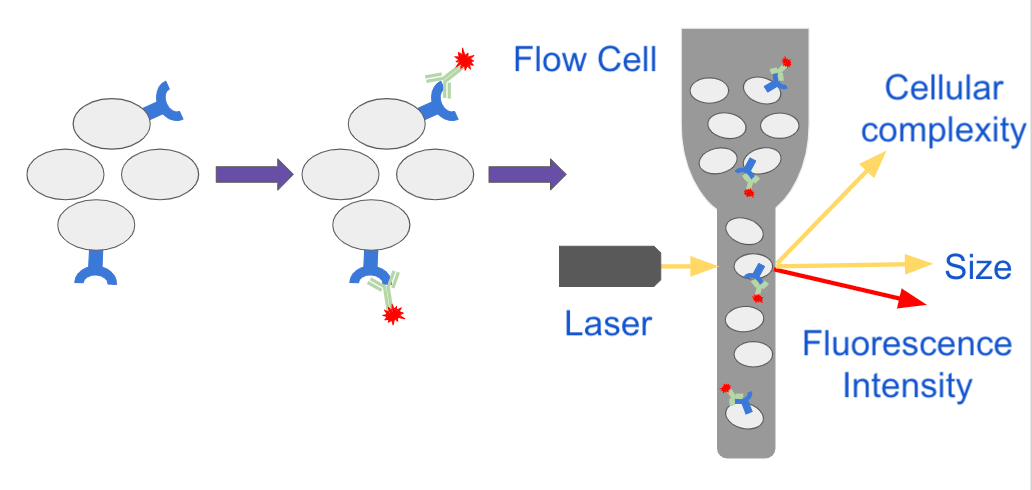
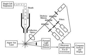
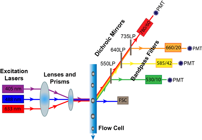
This helps ensure that every cell is analyzed independently. Its working depends on the light scattering features of the cells under investigation which may be derived from dyes or monoclonal antibodies targeting either extracellular molecules. Flow cytometry is a technique that employs an optical-electronic detection apparatus to analyze the physical and chemical properties of microscopic particles.
Flow cytometry provides a well-established method to identify cells in solution and is most commonly used for evaluating peripheral blood bone marrow and other body fluids. It has the functionality of standard flow cytometry and generates highresolution digital images of each. In flow cytometry each detection event cell is associated with several numerical measurements of fluorescence intensity and the degree of forward and side scatter of laser light.
Measurement of forward and side scatter of light Cells or particles passing through the beam scatter light which is detected as FS and SS. With the combined advantages of optical microscopy and flow cytometry imaging flow cytometry IFC has quickly become an established tool for performing cytometric analysis in diverse areas of biology including microbiology immunology and stem cell biology114. Compared with traditional flow cytometry imaging flow cytometry increases the number of parameters one can measure by providing morphological and spatial information in a high-throughput controlled manner.
Flow cytometry is a sophisticated instrument measuring multiple physical characteristics of a single cell such as size and granularity simultaneously as the cell flows in suspension through a measuring device. Imaging flow cytometry is emerging as a diagnostic tool for the assessment of leukemia. In Flow Cytometry Image Is Everything.
Cytometry in its purest form is the measurement of cell characteristics which can include cell size cell count cell cycle and more. The most critical requirement for efficient and effective flow cytometry analysis is that the sample be a single-cell suspension. Forward scatter is roughly correlated to the size of the cell and side scatter gives an indication of the cells granularity but flow cytometry.
Deep learning With the combined advantages of optical microscopy and flow cytometry imaging flow cytometry IFC has quickly become an established tool for performing cytometric. While non-image-based Flow Cytometry NIFC is a powerful multi-parameter high-throughput cytometric technology with widespread applications it cannot provide the spatialmorphometric information often essential to addressing key biological questions. The sample must therefore be ordered into a stream of single particles that can be.
The more recent expansion of flow. Flow cytometry is the measurement of cell characteristics which can include cell size cell count cell cycle and more. Imaging flow cytometry does just that.
One of the biggest differences between an imaging cytometer and a flow cytometer is that the former images. Cells set up for imaging cytometry are either stained to enhance contrast or are stained with antibodies directly conjugated to fluorophores. This flow cytometry guide aims to give you a basic overview of all the important aspects of flow cytometry.
1Department of Chemistry University of Tokyo Tokyo Japan. Thus Leeuwenhoek is often cited in any discussion. Flow Cytometry is the technical process that allows for the individual measurements of cell fluorescence and light scattering.
When a sample enters a flow cytometer the particles are randomly distributed in the 3-D space of the sample line the diameter of which is significantly larger than the diameter of most cells.

Representative Flow Cytometric Cytogram And Histogram A Cytogram Of Download Scientific Diagram
How Does Flow Cytometry Work Nanocellect

Resolution Of Flow Cytometry Data What It Means And Why It Matters For Publications Marissa Fahlberg Phd
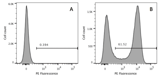
Putting Down A Marker In Flow Cytometry To Help Determine Positivity

Introduction To Flow Cytometry Flow Cytometry Medical Technology Medical Laboratory Science

Flow Cytometry Measurements For Chlorella Vulgaris Cells Labeled With Download Scientific Diagram
Analyzing Flow Cytometry Results Nanocellect
Flow Cytometry Archives Bitesize Bio
How Does Flow Cytometry Work Nanocellect

How To Identify Bad Flow Cytometry Data Bad Data Part 1 Cytometry And Antibody Technology
Overview Of The Flow Cytometer Flow Cytometry Flow Medical Laboratory Science

How To Identify Bad Flow Cytometry Data Bad Data Part 1 Cytometry And Antibody Technology

Flow Cytometry Vector Illustration Flow Cytometry Vector Illustration Illustration

Diagnostic Potential Of Imaging Flow Cytometry Trends In Biotechnology

Flow Cytometry And The Sheath Fluid You Use Lab Manager

Flow Cytometry Definition Principle Parts Steps Types Uses Flow Cytometry Principles Flow

Putting Down A Marker In Flow Cytometry To Help Determine Positivity

Flow Cytometry Antibodies Tips Flow Cytometry Flow Life Science

Resolution Of Flow Cytometry Data What It Means And Why It Matters For Publications Immunowiz


0 komentar:
Posting Komentar